
1-6 on Lees morning chest x-ray it showed that the stent had moved into his stomach, he was taken back to endoscopy to have it adjusted.
1-7 the stent once again moved into his stomach over night so the plan is to remove the stent and replace the feeding tube. this will happen on 1-8.
With the stent failing it is time to figure something out to fix Lees achalasia. There are two invasive procedures that are the treatment for it.
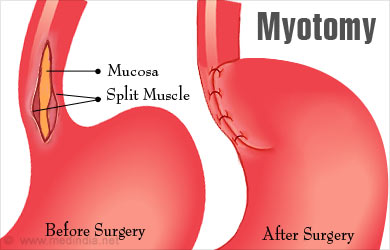
First there is the Heller myotomy.
The Heller myotomy is a laparoscopic (minimally invasive) surgical procedure used to treat achalasia. Achalasia is a disorder of the esophagus that makes it hard for foods and liquids to pass into the stomach.The Heller myotomy is essentially an esophagomyotomy, the cutting the esophageal sphincter muscle, performed laparoscopically.
This surgery comes with risk to Lee with his liver failure it increase the risks to be a high risk surgery. There are so many factors that can make the surgery high risk the risk for infection is higher due to the ascities, risk of not tolerating anesthesia, and just general risks of making the liver failure worse.
The second invasive procedure is a balloon dilation.

The balloon dilation also comes with risk. There is a risk of rupturing the esophagus and the risk of bleeding.
There are risks with both procedures but the liver team has decided that the balloon dilation has the less risks. There are a couple of things that will be done to decrease the risk before the balloon dilation. The team will set Lee up to have a TIPS procedure and have the varices in his esophagus embolized.
TIPS is a non-surgical method of placing a portosystemic shunt. The shunt is passed down the jugular vein from the neck by a radiologist using X-ray guidance. The shunt then is inserted between the portal and hepatic veins within the liver.

On the way home we were at a stand still on the interstate, it took a couple hours to get home. Lee had a good nap. There was a accident that had traffic shut down to a stand still.


No comments:
Post a Comment